Artículos de investigación
Estos artículos están dirigidos a los médicos y la comunidad científica. Muchos de ellos se encuentran indexados en la Biblioteca Nacional de Medicina de Estados Unidos (en inglés National Library of Medicine o NLM).
Si desea consultar acerca de los temas tratados puede agendar una cita con el Dr. Cruzado o escribir al correo electrónico en el área de contacto.
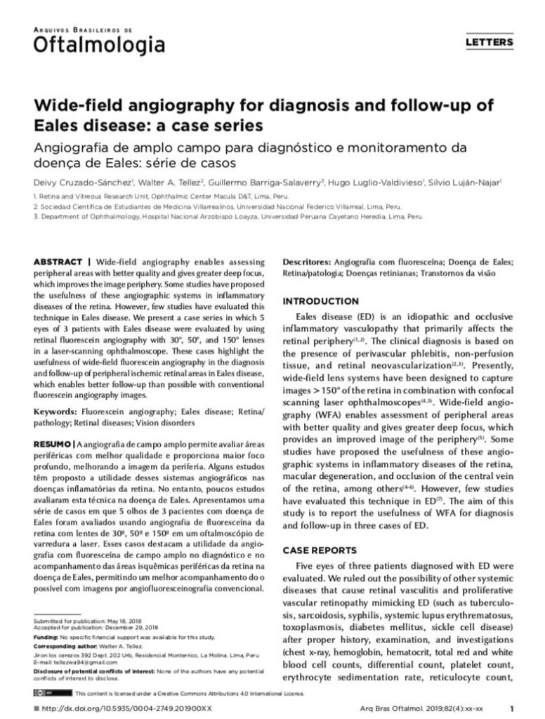
Wide-field angiography for diagnosis and follow-up of Eales disease: a case series.
Cruzado-Sánchez D1, Tellez WA2, Barriga-Salaverry G3, Luglio-Valdivieso H1, Luján-Najar S1.
Abstract
Wide-field angiography enables assessing peripheral areas with better quality and gives greater deep focus, which improves the image periphery. Some studies have proposed the usefulness of these angiographic systems in inflammatory diseases of the retina. However, few studies have evaluated this technique in Eales disease. We present a case series in which 5 eyes of 3 patients with Eales disease were evaluated by using retinal fluorescein angiography with 30º, 50º, and 150º lenses in a laser-scanning ophthalmoscope. These cases highlight the usefulness of wide-field fluorescein angiography in the diagnosis and follow-up of peripheral ischemic retinal areas in Eales disease, which enables better follow-up than possible with conventional fluorescein angiography images.
PMID: 31116318
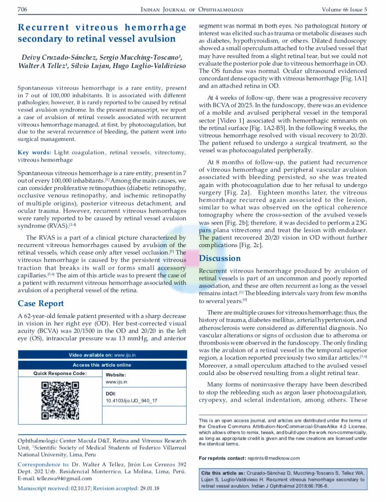
Recurrent vitreous hemorrhage secondary to retinal vessel avulsion.
Cruzado-Sanchez D1, Mucching-Toscano S2, Tellez WA2, Lujan S1, Luglio-Valdivieso H1.
Abstract
Spontaneous vitreous hemorrhage is a rare entity, present in 7 out of 100,000 inhabitants. It is associated with different pathologies; however, it is rarely reported to be caused by retinal vessel avulsion syndrome. In the present manuscript, we report a case of avulsion of retinal vessels associated with recurrent vitreous hemorrhage managed, at first, by photocoagulation, but due to the several recurrence of bleeding, the patient went into surgical management.
KEYWORDS: Light coagulation; retinal vessels; vitrectomy; vitreous hemorrhage
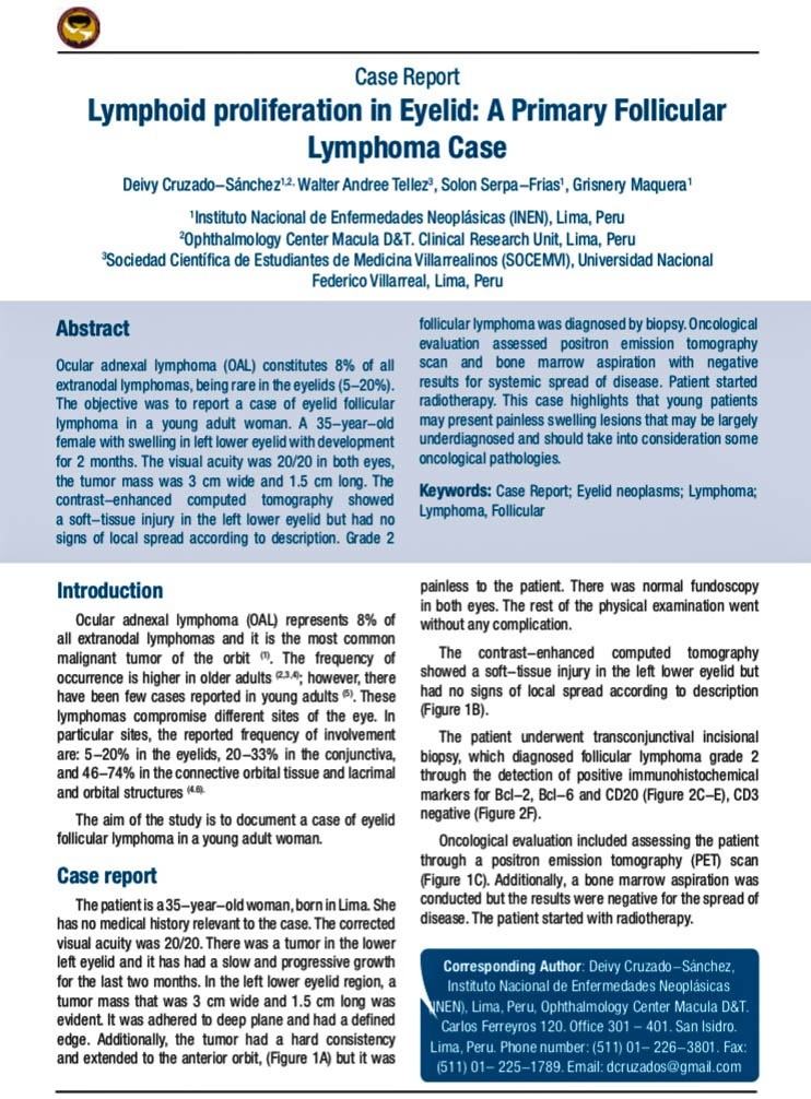
Lymphoid proliferation in Eyelid: A Primary Follicular Lymphoma Case.
Cruzado-Sánchez D1,2, Tellez WA3, Serpa-Frias S1, Maquera G1.
Abstract
Ocular adnexal lymphoma (OAL) constitutes 8% of all extranodal lymphomas, being rare in the eyelids (5-20%). The objective was to report a case of eyelid follicular lymphoma in a young adult woman. A 35-year-old female with swelling in left lower eyelid with development for 2 months. The visual acuity was 20/20 in both eyes, the tumor mass was 3 cm wide and 1.5 cm long. The contrast-enhanced computed tomography showed a soft-tissue injury in the left lower eyelid but had no signs of local spread according to description. Grade 2 follicular lymphoma was diagnosed by biopsy. Oncological evaluation assessed positron emission tomography scan and bone marrow aspiration with negative results for systemic spread of disease. Patient started radiotherapy. This case highlights that young patients may present painless swelling lesions that may be largely underdiagnosed and should take into consideration some oncological pathologies.
PMID: 29607824
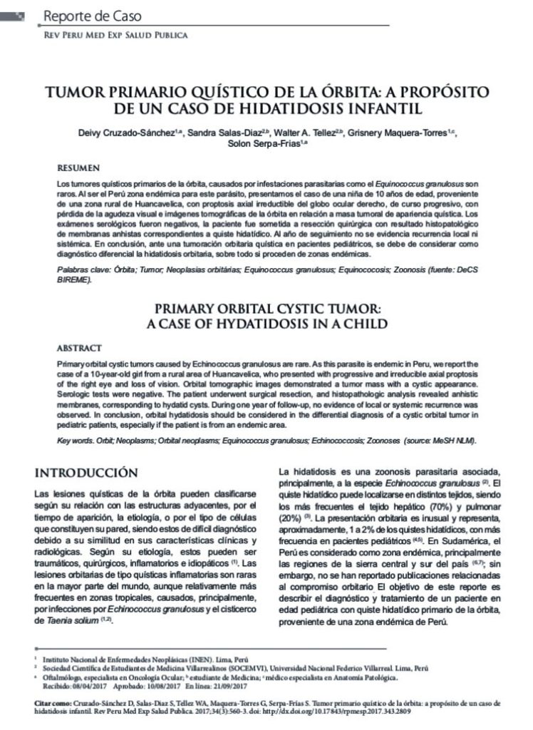
[Primary orbital cystic tumor: A case of hydatidosis in a child].
Cruzado-Sánchez D1, Salas-Diaz S2, Tellez WA2, Maquera-Torres G1, Serpa-Frias S1.
Abstract
Primary orbital cystic tumors caused by Echinococcus granulosus are rare. As this parasite is endemic in Peru, we report the case of a 10-year-old girl from a rural area of Huancavelica, who presented with progressive and irreducible axial proptosis of the right eye and loss of vision. Orbital tomographic images demonstrated a tumor mass with a cystic appearance. Serologic tests were negative. The patient underwent surgical resection, and histopathologic analysis revealed anhistic membranes, corresponding to hydatid cysts. During one year of follow-up, no evidence of local or systemic recurrence was observed. In conclusion, orbital hydatidosis should be considered in the differential diagnosis of a cystic orbital tumor in pediatric patients, especially if the patient is from an endemic area.
Interferon alpha-2a as alternative treatment for conjunctival squamous cell carcinoma.
Cruzado-Sánchez D1, Salas-Diaz M2, Tellez WA3, Alvarez-Matos SE3, Serpa-Frías S4.
Abstract
CASE REPORT:
A 35 year-old male patient with a history of HIV infection characterized by progressive tumour growth in bulbar conjunctiva of the left eye, corresponding to conjunctival squamous cell carcinoma that responded to treatment with interferon alpha-2a.
DISCUSSION:
Interferon alpha-2b has been used at conjunctival level as a topical immunomodulator treatment, with complete remission of epithelial neoplasms being observed. However, there have not been any previous publications on the use of interferon alpha-2a, which differs from interferon alpha-2b in a single amino acid, for the treatment of conjunctival squamous cell carcinoma.
KEYWORDS:
Carcinoma conjuntival de células escamosas; Conjunctiva; Conjunctival squamous cell carcinoma; Conjuntiva; Interferon alpha-2a; Interferón alfa-2ª
Cruzado-Sánchez D, Luglio Valdivieso H, Lujan Nájar SM.
[Indexed for MEDLINE]
Cruzado-Sánchez D1, Tobón C, Lujan V, Lujan S, Valderrama V
Abstract
The case of a 36 year-old male with neuroretinitis caused by Bartonella henselae is reported, whose periodic follow-up was done through optical coherence tomography (OCT). The onset of this disease was characterized by unilateral low visual acuity (VA), painless, of sudden onset, in the right eye (RE), associated to l febrile symptom. The funduscopic examination showed edema in the posterior pole which extended from the optical disc to the macular region in the RE. The OCT confirmed macular and optical disc thickening, as well as the presence of subretinal macular fluid. Systemic studies were normal except for a blood count due to the presence of leukocytosis and positive for Bartonella henselae. The follow up with CT Scan helped to evaluate the decrease in macular edema, with the subsequent improvement of visual acuity and absence of related complications. This report describes the utility of the follow up with OCT in a patient with neuroretinitis caused by Bartonella henselae.
[Indexed for MEDLINE]
Cruzado-Sánchez D; Tobón C; Luglio H; Lujan S.
Abstract
Objectives: Describe the images of the choroid by optical coherence tomography (OCT) spectral domain in healthy subjects and chorioretinal diseases.
Materials and methods:
Case series study. We evaluated 67 cases involving healthy ocular subjects and patients with chorioretinal diseases. The scans included a sweep of the retina and choroid at level of the macula conventionally, and other using enhanced depth imaging (EDI). The choroid thickness was measured from the outer edge of the retinal pigment epithelium to the internal scleral rim.
Results: Of the 67 cases, 12 (17.4%) were healthy ocular subjects, 10 (14.5%) with central serous chorioretinopathy (CSC), 23 (33.4%) agerelated macular degeneration (AMD), 9 (13.0%) with myopia, 13 (18.8%) with polypoidal choroidal vasculopathy. There are differences in the quality of images with conventional OCT and those with EDI, allowing only to describe the choriocapillaris in some cases while the EDI protocol can appreciate the total thickness of the choroid. The choroid is thicker in polypoidal choroidal vasculopathy (353.92 ± 68.66 microns) and CSC (408.81 ± 95.56 microns) than in healthy subjects (251.97 ± 66.37 mm) (p<0.05) and is decreased in the dry AMD (124.69 ± 34.02 microns) (p<0.05), as well as in the myopia (118.47 ± 34.02 microns) (p<0.05).
Conclusions: OCT images of the choroid by PIM is a reproducible technique for describing and measuring its thickness, being decreased in AMD and myopia, and was increased in the polypoidal vasculopathy and CSC.
Endoscopic Surgical Management of Dacryocele.
Morante, J. C. C., Venegas, M. M. H., Morante, A. C., Valdivia, V., Cruzado-Sanchez, D., & Chang, J. H. (2013).
Abstract
Objectives: 1) Report our experience and surgical treatment of the unilateral giant dacryocele using the endoscopic technique. 2) Improve the lacrimal sac treatment with endonasal surgery, thus avoiding external dacryocystorhinostomy and its sequelae.
Methods: We present the case of an adult patient with chronic dacryocystitis of 1 year of evolution, treated by endoscopic endonasal surgery (endoscopic dacryocystorhinostomy) in July 2012 at the Arzobispo Loayza National Hospital, Lima, Peru. The computed tomography described a giant mucocele of the right lacrimal sac. The technique was done entirely endoscopically, opening the lacrimal bone and the lacrimal sac with mucosal content and using a silicone probe. Nasal packing was placed for 3 days, and endoscopic control was carried out.
Results: This patient was followed endoscopically each week, testing the permeability of the fistula, moving the probe, and checking the opening hole during the 6 months after surgery. There were no recurrences and no complications.
Conclusions: Endoscopic dacryocystorhinostomy has become a very safe method to resolve lacrimal sac pathologies. It is a reliable and secure and has less sequelae than external dacryocystorhinostomies.
Venegas, M. M. H., Morante, J. C. C., Cruzado-Sanchez, D., Valdivia, V., & Chang, J. H.
Abstract
Objectives: Describe the complications of endoscopic and conventional dacryocystorhinostomy (external) in chronic dacryocystitis (CDC) in a public hospital.
Methods: Case series. We evaluated 39 patients who were treated surgically of CDC by endoscopic or conventional dacryocystorhinostomy between January 2006 and December 2011 at the Arzobispo Loayza National Hospital, Lima,Peru. We evaluated the complications associated with the treatment received.
Results: Of the 39cases, 29 (74.4%) were women, and the average age was 54.9 years. The mean disease duration was 29.7 months. The affected side was the right in 59%, and the most common symptom was epiphora (84.6%), followed by secretion (7.7%) and tumor (5.1%). 56.4% of the surgeries were made by conventional technique. The most common complication was the post-surgical removal of the probe in 4 patients, 3 (7.7%) by the conventional and 1 (2.5%) by the endoscopical technique. Other complications were reocclusion (5.2%), abscess (2.6%), and epistaxis (2.6%), all for the conventional technique. By the endoscopical technique, just the infection (5.2%) and synechiae (2.6%).
Conclusions: Complications associated with the conventional technique were expulsion of the probe (7.7%), reocclusion (5.2%), abscess (2.6%), and epistaxis (2.6%), while complications with the endoscopic technique were removal of the probe (2.5 %), infection (5.2%), and synechiae (2.6%)
Factores de riesgo neonatales asociados a retinopatía de la prematuridad.
Díaz M; Cruzado-Sánchez D.
Resumen
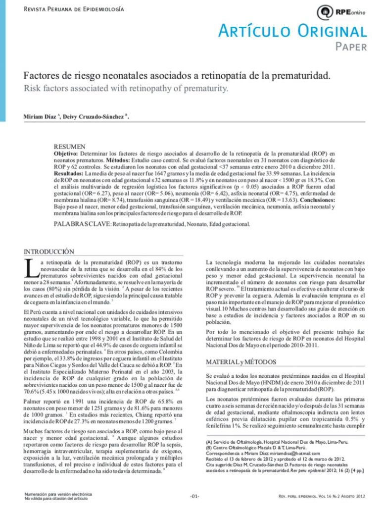
Objetivo: Determinar los factores de riesgo asociados al desarrollo de la retinopatía de la prematuridad (ROP) en neonatos prematuros. Método: Estudio caso control. Se evaluó factores neonatales en 31 neonatos con diagnóstico de ROP y 62 controles. Se estudiaron los neonatos con edad gestacional <37 semanas entre enero 2010 a diciembre 2011. Resultados: La media de peso al nacer fue 1647 gramos y la media de edad gestacional fue 33.99 semanas. La incidencia de ROP en neonatos con edad gestacional 32 semanas es 11.8%y en neonatos con peso al nacer< 1500 gr es la.3%. Con el análisis multivariado de regresión logística los :factores significativos (p < 0.05) asociadas a ROP fueron edad gestacional (OR- ó27), peso al nacer (OR=- 5.0ó), neumonía (OR=- ó.42), asfixia neonatal (OR- 4.75), enfermedad de membrana hialina(OR- 8.74), transfusión sanguínea (OR=18.49) ventilación mecánica(OR- 13.ó3). Conclusiones: Bajo peso al nacer, menor edad gestacional, transfusión sanguínea, ventilación mecánica, neumonía, asfixia neonatal y membrana hialina son los principales factores de riesgo para el desarrollo de ROP.
Palabras clave: Retinopatía de la prematuridad, Neonato, Edad gestacional.
Valdez JA, Cruzado -Sánchez D.
Resumen
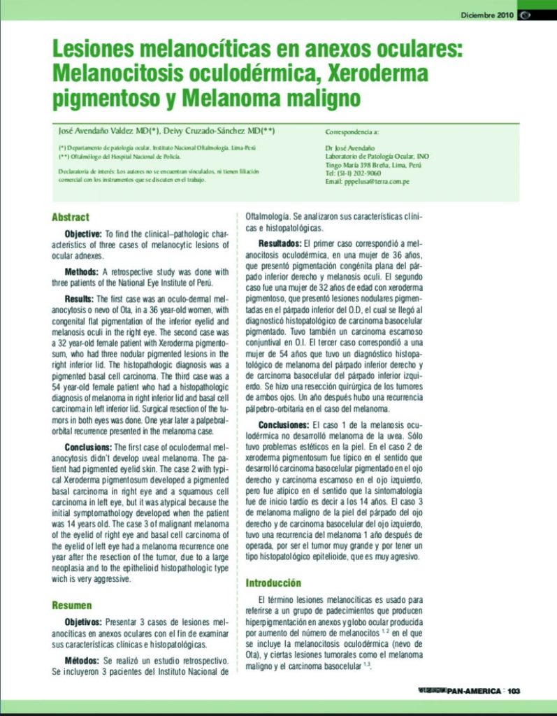
Objetivos: Presentar 3 casos de lesiones melanocíticas en anexos oculares con el fin de examinar sus características clínicas e histopatológicas. Métodos: Se realizó un estudio retrospectivo. Se incluyeron 3 pacientes del Instituto Nacional de Oftalmología. Se analizaron sus características clíni-cas e histopatológicas. Resultados: El primer caso correspondió a mel-anocitosis oculodérmica, en una mujer de 36 años, que presentó pigmentación congénita plana del pár-pado inferior derecho y melanosis oculi. El segundo caso fue una mujer de 32 años de edad con xeroderma pigmentoso, que presentó lesiones nodulares pigmen-tadas en el párpado inferior del O.D, el cual se llegó al diagnosticó histopatológico de carcinoma basocelular pigmentado. Tuvo también un carcinoma escamoso conjuntival en O.I. El tercer caso correspondió a una mujer de 54 años que tuvo un diagnóstico histopa-tológico de melanoma del párpado inferior derecho y de carcinoma basocelular del párpado inferior izquierdo. Se hizo una resección quirúrgica de los tumores de ambos ojos. Un año después hubo una recurrencia pálpebro-orbitaria en el caso del melanoma. Conclusiones: El caso 1 de la melanosis ocu-lodérmica no desarrolló melanoma de la uvea. Sólo tuvo problemas estéticos en la piel. En el caso 2 de xeroderma pigmentosum fue típico en el sentido que desarrolló carcinoma basocelular pigmentado en el ojo derecho y carcinoma escamoso en el ojo izquierdo, pero fue atípico en el sentido que la sintomatología fue de inicio tardío es decir a los 14 años. El caso 3 de melanoma maligno de la piel del párpado del ojo derecho y de carcinoma basocelular del ojo izquierdo, tuvo una recurrencia del melanoma 1 año después de operada, por ser el tumor muy grande y por tener un tipo histopatológico epitelioide, que es muy agresivo.
Cruzado-Sánchez, D. Bedriñana-Arones, L. Mayta-Salinas, E. Cupe-Chacalcaje, K. Álvarez-Romero, F. Sánchez-Malpica, M.
Resumen
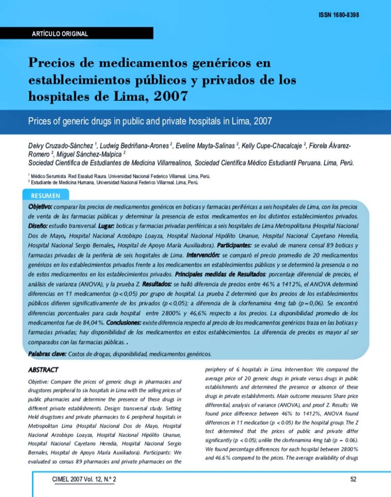
Objetivo: comparar los precios de medicamentos genéricos en boticas y farmacias periféricas a seis hospitales de Lima, con los precios de venta de las farmacias públicas y determinar la presencia de estos medicamentos en los distintos establecimientos privados.
Diseño: estudio transversal. Lugar: boticas y farmacias privadas periféricas a seis hospitales de Lima Metropolitana (Hospital Nacional Dos de Mayo, Hospital Nacional Arzobispo Loayza, Hospital Nacional Hipólito Unanue, Hospital Nacional Cayetano Heredia, Hospital Nacional Sergio Bernales, Hospital de Apoyo María Auxiliadora). Participantes: se evaluó de manera censal 89 boticas y farmacias privadas de la periferia de seis hospitales de Lima. Intervención: se comparó el precio promedio de 20 medicamentos
genéricos en los establecimientos privados frente a los medicamentos en establecimientos públicos y se determinó la presencia o no de estos medicamentos en los establecimientos privados. Principales medidas de Resultados: porcentaje diferencial de precios, el análisis de varianza (ANOVA), y la prueba Z. Resultados: se halló diferencia de precios entre 46% a 1412%, el ANOVA determinó diferencias en 11 medicamentos (p<0,05) por grupo de hospital. La prueba Z determinó que los precios de los establecimientos públicos difieren significativamente de los privados (p<0,05); a diferencia de la clorfenamina 4mg tab (p=0,06). Se encontró diferencias porcentuales para cada hospital entre 2800% y 46,6% respecto a los precios. La disponibilidad promedio de los medicamentos fue de 84,04%. Conclusiones: existe diferencia respecto al precio de los medicamentos genéricos traza en las boticas y farmacias privadas; hay disponibilidad de los medicamentos en estos establecimientos. La diferencia de precios es mayor al ser comparados con las farmacias públicas.
Palabras clave: Costos de drogas, disponibilidad, medicamentos genéricos.
Espirometría forzada en pobladores de alturas expuestos al humo de biomasas y su asociación con EPOC
Cruzado-Sanchez D; Guerrero R; Hinostroza L.
Resumen
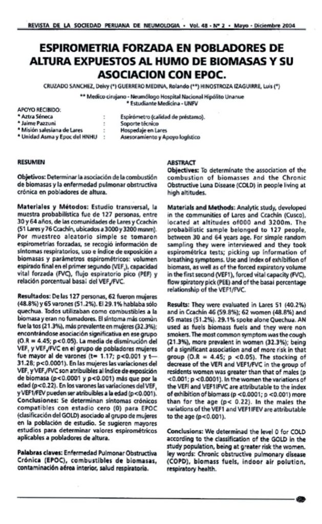
Objetivos: Determinar la asociación de la combustión de biomasas y la enfermedad pulmonar obstructiva crónica en pobladores de altura. Materiales y Métodos: Estudio transversal, la muestra probabilística fue de 127 personas, entre 30 y 64 años, de las comunidades de lares y Ccachín (51 la resy76 Ccachín, ubicados a 3000 y 3200 msnm). Por muestreo aleatorio simple se tomaron espirometrías forzadas, se recogió información de síntomas respiratorios, uso e índice de exposición a biomasas y parßmetros espiro métricos: volumen espirado final en el primer segundo (VEF,), capacidad vital forzada (FVC), flujo espiratorio pico (PEF) y relación porcentual basai del VEF, IFVC. Resultados: De las 127 personas, 62 fueron mujeres (48.8 por ciento) y 65 varones (51.2 por ciento ). El 29.1 por ciento hablaba sólo quechua. Todos utilizaban como combustibles a la biomasa y eran no fumadores. El síntoma mßs común fue la tos (21.3 por ciento ), mßs prevalente en mujeres (32.3 por ciento); encontrßndose asociación significativa en ese grupo (O.R= 4.45; p menor que 0.05). La media de disminución del VEF1, y VEF1/FVC en el grupo de pobladores mujeres fue mayor al de varones (t= 1.17; p menor que 0.001 y t- 31.28; p menor que 0.0001). En las mujeres las variaciones del VEF1,y VEF1/FVC son atribuibles al índice de exposición de biomasa (p menor que 0.0001 y p menor que 0.001) mßs que por la edad (p menor que 0.22). En los varones las variaciones del VEF1, y VEF1/FEV pueden ser atribuibles a la edad (p menor que 0.001). Conclusiones: Se determinan síntomas crónicos compatibles con estadio cero (O) para EPOC (clasificación del GOLD) asociado al grupo de mujeres en la población de estudio. Se sugieren mayores estudios para determinar valores espirométricos aplicables a pobladores de altura.
Artículos de investigación
Estos artículos están dirigidos a los médicos y la comunidad científica. Muchos de ellos se encuentran indexados en la Biblioteca Nacional de Medicina de Estados Unidos (en inglés National Library of Medicine o NLM).
Si desea consultar acerca de los temas tratados puede agendar una cita con el Dr. Cruzado o escribir al correo electrónico en el área de contacto.
Wide-field angiography for diagnosis and follow-up of Eales disease: a case series.
Cruzado-Sánchez D1, Tellez WA2, Barriga-Salaverry G3, Luglio-Valdivieso H1, Luján-Najar S1.
Abstract
Wide-field angiography enables assessing peripheral areas with better quality and gives greater deep focus, which improves the image periphery. Some studies have proposed the usefulness of these angiographic systems in inflammatory diseases of the retina. However, few studies have evaluated this technique in Eales disease. We present a case series in which 5 eyes of 3 patients with Eales disease were evaluated by using retinal fluorescein angiography with 30º, 50º, and 150º lenses in a laser-scanning ophthalmoscope. These cases highlight the usefulness of wide-field fluorescein angiography in the diagnosis and follow-up of peripheral ischemic retinal areas in Eales disease, which enables better follow-up than possible with conventional fluorescein angiography images.
PMID: 31116318
Recurrent vitreous hemorrhage secondary to retinal vessel avulsion.
Cruzado-Sanchez D1, Mucching-Toscano S2, Tellez WA2, Lujan S1, Luglio-Valdivieso H1.
Abstract
Spontaneous vitreous hemorrhage is a rare entity, present in 7 out of 100,000 inhabitants. It is associated with different pathologies; however, it is rarely reported to be caused by retinal vessel avulsion syndrome. In the present manuscript, we report a case of avulsion of retinal vessels associated with recurrent vitreous hemorrhage managed, at first, by photocoagulation, but due to the several recurrence of bleeding, the patient went into surgical management.
KEYWORDS: Light coagulation; retinal vessels; vitrectomy; vitreous hemorrhage
Lymphoid proliferation in Eyelid: A Primary Follicular Lymphoma Case.
Cruzado-Sánchez D1,2, Tellez WA3, Serpa-Frias S1, Maquera G1.
Abstract
Ocular adnexal lymphoma (OAL) constitutes 8% of all extranodal lymphomas, being rare in the eyelids (5-20%). The objective was to report a case of eyelid follicular lymphoma in a young adult woman. A 35-year-old female with swelling in left lower eyelid with development for 2 months. The visual acuity was 20/20 in both eyes, the tumor mass was 3 cm wide and 1.5 cm long. The contrast-enhanced computed tomography showed a soft-tissue injury in the left lower eyelid but had no signs of local spread according to description. Grade 2 follicular lymphoma was diagnosed by biopsy. Oncological evaluation assessed positron emission tomography scan and bone marrow aspiration with negative results for systemic spread of disease. Patient started radiotherapy. This case highlights that young patients may present painless swelling lesions that may be largely underdiagnosed and should take into consideration some oncological pathologies.
PMID: 29607824
[Primary orbital cystic tumor: A case of hydatidosis in a child].
Cruzado-Sánchez D1, Salas-Diaz S2, Tellez WA2, Maquera-Torres G1, Serpa-Frias S1.
Abstract
Primary orbital cystic tumors caused by Echinococcus granulosus are rare. As this parasite is endemic in Peru, we report the case of a 10-year-old girl from a rural area of Huancavelica, who presented with progressive and irreducible axial proptosis of the right eye and loss of vision. Orbital tomographic images demonstrated a tumor mass with a cystic appearance. Serologic tests were negative. The patient underwent surgical resection, and histopathologic analysis revealed anhistic membranes, corresponding to hydatid cysts. During one year of follow-up, no evidence of local or systemic recurrence was observed. In conclusion, orbital hydatidosis should be considered in the differential diagnosis of a cystic orbital tumor in pediatric patients, especially if the patient is from an endemic area.
Interferon alpha-2a as alternative treatment for conjunctival squamous cell carcinoma.
Cruzado-Sánchez D1, Salas-Diaz M2, Tellez WA3, Alvarez-Matos SE3, Serpa-Frías S4.
Abstract
CASE REPORT:
A 35 year-old male patient with a history of HIV infection characterized by progressive tumour growth in bulbar conjunctiva of the left eye, corresponding to conjunctival squamous cell carcinoma that responded to treatment with interferon alpha-2a.
DISCUSSION:
Interferon alpha-2b has been used at conjunctival level as a topical immunomodulator treatment, with complete remission of epithelial neoplasms being observed. However, there have not been any previous publications on the use of interferon alpha-2a, which differs from interferon alpha-2b in a single amino acid, for the treatment of conjunctival squamous cell carcinoma.
KEYWORDS:
Carcinoma conjuntival de células escamosas; Conjunctiva; Conjunctival squamous cell carcinoma; Conjuntiva; Interferon alpha-2a; Interferón alfa-2ª
Cruzado-Sánchez D, Luglio Valdivieso H, Lujan Nájar SM.
[Indexed for MEDLINE]
Cruzado-Sánchez D1, Tobón C, Lujan V, Lujan S, Valderrama V
Abstract
The case of a 36 year-old male with neuroretinitis caused by Bartonella henselae is reported, whose periodic follow-up was done through optical coherence tomography (OCT). The onset of this disease was characterized by unilateral low visual acuity (VA), painless, of sudden onset, in the right eye (RE), associated to l febrile symptom. The funduscopic examination showed edema in the posterior pole which extended from the optical disc to the macular region in the RE. The OCT confirmed macular and optical disc thickening, as well as the presence of subretinal macular fluid. Systemic studies were normal except for a blood count due to the presence of leukocytosis and positive for Bartonella henselae. The follow up with CT Scan helped to evaluate the decrease in macular edema, with the subsequent improvement of visual acuity and absence of related complications. This report describes the utility of the follow up with OCT in a patient with neuroretinitis caused by Bartonella henselae.
[Indexed for MEDLINE]
Cruzado-Sánchez D; Tobón C; Luglio H; Lujan S.
Abstract
Objectives: Describe the images of the choroid by optical coherence tomography (OCT) spectral domain in healthy subjects and chorioretinal diseases.
Materials and methods:
Case series study. We evaluated 67 cases involving healthy ocular subjects and patients with chorioretinal diseases. The scans included a sweep of the retina and choroid at level of the macula conventionally, and other using enhanced depth imaging (EDI). The choroid thickness was measured from the outer edge of the retinal pigment epithelium to the internal scleral rim.
Results: Of the 67 cases, 12 (17.4%) were healthy ocular subjects, 10 (14.5%) with central serous chorioretinopathy (CSC), 23 (33.4%) agerelated macular degeneration (AMD), 9 (13.0%) with myopia, 13 (18.8%) with polypoidal choroidal vasculopathy. There are differences in the quality of images with conventional OCT and those with EDI, allowing only to describe the choriocapillaris in some cases while the EDI protocol can appreciate the total thickness of the choroid. The choroid is thicker in polypoidal choroidal vasculopathy (353.92 ± 68.66 microns) and CSC (408.81 ± 95.56 microns) than in healthy subjects (251.97 ± 66.37 mm) (p<0.05) and is decreased in the dry AMD (124.69 ± 34.02 microns) (p<0.05), as well as in the myopia (118.47 ± 34.02 microns) (p<0.05).
Conclusions: OCT images of the choroid by PIM is a reproducible technique for describing and measuring its thickness, being decreased in AMD and myopia, and was increased in the polypoidal vasculopathy and CSC.
Endoscopic Surgical Management of Dacryocele.
Morante, J. C. C., Venegas, M. M. H., Morante, A. C., Valdivia, V., Cruzado-Sanchez, D., & Chang, J. H. (2013).
Abstract
Objectives: 1) Report our experience and surgical treatment of the unilateral giant dacryocele using the endoscopic technique. 2) Improve the lacrimal sac treatment with endonasal surgery, thus avoiding external dacryocystorhinostomy and its sequelae.
Methods: We present the case of an adult patient with chronic dacryocystitis of 1 year of evolution, treated by endoscopic endonasal surgery (endoscopic dacryocystorhinostomy) in July 2012 at the Arzobispo Loayza National Hospital, Lima, Peru. The computed tomography described a giant mucocele of the right lacrimal sac. The technique was done entirely endoscopically, opening the lacrimal bone and the lacrimal sac with mucosal content and using a silicone probe. Nasal packing was placed for 3 days, and endoscopic control was carried out.
Results: This patient was followed endoscopically each week, testing the permeability of the fistula, moving the probe, and checking the opening hole during the 6 months after surgery. There were no recurrences and no complications.
Conclusions: Endoscopic dacryocystorhinostomy has become a very safe method to resolve lacrimal sac pathologies. It is a reliable and secure and has less sequelae than external dacryocystorhinostomies.
Venegas, M. M. H., Morante, J. C. C., Cruzado-Sanchez, D., Valdivia, V., & Chang, J. H.
Abstract
Objectives: Describe the complications of endoscopic and conventional dacryocystorhinostomy (external) in chronic dacryocystitis (CDC) in a public hospital.
Methods: Case series. We evaluated 39 patients who were treated surgically of CDC by endoscopic or conventional dacryocystorhinostomy between January 2006 and December 2011 at the Arzobispo Loayza National Hospital, Lima,Peru. We evaluated the complications associated with the treatment received.
Results: Of the 39cases, 29 (74.4%) were women, and the average age was 54.9 years. The mean disease duration was 29.7 months. The affected side was the right in 59%, and the most common symptom was epiphora (84.6%), followed by secretion (7.7%) and tumor (5.1%). 56.4% of the surgeries were made by conventional technique. The most common complication was the post-surgical removal of the probe in 4 patients, 3 (7.7%) by the conventional and 1 (2.5%) by the endoscopical technique. Other complications were reocclusion (5.2%), abscess (2.6%), and epistaxis (2.6%), all for the conventional technique. By the endoscopical technique, just the infection (5.2%) and synechiae (2.6%).
Conclusions: Complications associated with the conventional technique were expulsion of the probe (7.7%), reocclusion (5.2%), abscess (2.6%), and epistaxis (2.6%), while complications with the endoscopic technique were removal of the probe (2.5 %), infection (5.2%), and synechiae (2.6%)
Factores de riesgo neonatales asociados a retinopatía de la prematuridad.
Díaz M; Cruzado-Sánchez D.
Resumen
Objetivo: Determinar los factores de riesgo asociados al desarrollo de la retinopatía de la prematuridad (ROP) en neonatos prematuros. Método: Estudio caso control. Se evaluó factores neonatales en 31 neonatos con diagnóstico de ROP y 62 controles. Se estudiaron los neonatos con edad gestacional <37 semanas entre enero 2010 a diciembre 2011. Resultados: La media de peso al nacer fue 1647 gramos y la media de edad gestacional fue 33.99 semanas. La incidencia de ROP en neonatos con edad gestacional 32 semanas es 11.8%y en neonatos con peso al nacer< 1500 gr es la.3%. Con el análisis multivariado de regresión logística los :factores significativos (p < 0.05) asociadas a ROP fueron edad gestacional (OR- ó27), peso al nacer (OR=- 5.0ó), neumonía (OR=- ó.42), asfixia neonatal (OR- 4.75), enfermedad de membrana hialina(OR- 8.74), transfusión sanguínea (OR=18.49) ventilación mecánica(OR- 13.ó3). Conclusiones: Bajo peso al nacer, menor edad gestacional, transfusión sanguínea, ventilación mecánica, neumonía, asfixia neonatal y membrana hialina son los principales factores de riesgo para el desarrollo de ROP.
Palabras clave: Retinopatía de la prematuridad, Neonato, Edad gestacional.
Valdez JA, Cruzado -Sánchez D.
Resumen
Objetivos: Presentar 3 casos de lesiones melanocíticas en anexos oculares con el fin de examinar sus características clínicas e histopatológicas. Métodos: Se realizó un estudio retrospectivo. Se incluyeron 3 pacientes del Instituto Nacional de Oftalmología. Se analizaron sus características clíni-cas e histopatológicas. Resultados: El primer caso correspondió a mel-anocitosis oculodérmica, en una mujer de 36 años, que presentó pigmentación congénita plana del pár-pado inferior derecho y melanosis oculi. El segundo caso fue una mujer de 32 años de edad con xeroderma pigmentoso, que presentó lesiones nodulares pigmen-tadas en el párpado inferior del O.D, el cual se llegó al diagnosticó histopatológico de carcinoma basocelular pigmentado. Tuvo también un carcinoma escamoso conjuntival en O.I. El tercer caso correspondió a una mujer de 54 años que tuvo un diagnóstico histopa-tológico de melanoma del párpado inferior derecho y de carcinoma basocelular del párpado inferior izquierdo. Se hizo una resección quirúrgica de los tumores de ambos ojos. Un año después hubo una recurrencia pálpebro-orbitaria en el caso del melanoma. Conclusiones: El caso 1 de la melanosis ocu-lodérmica no desarrolló melanoma de la uvea. Sólo tuvo problemas estéticos en la piel. En el caso 2 de xeroderma pigmentosum fue típico en el sentido que desarrolló carcinoma basocelular pigmentado en el ojo derecho y carcinoma escamoso en el ojo izquierdo, pero fue atípico en el sentido que la sintomatología fue de inicio tardío es decir a los 14 años. El caso 3 de melanoma maligno de la piel del párpado del ojo derecho y de carcinoma basocelular del ojo izquierdo, tuvo una recurrencia del melanoma 1 año después de operada, por ser el tumor muy grande y por tener un tipo histopatológico epitelioide, que es muy agresivo.
Cruzado-Sánchez, D. Bedriñana-Arones, L. Mayta-Salinas, E. Cupe-Chacalcaje, K. Álvarez-Romero, F. Sánchez-Malpica, M.
Resumen
Objetivo: comparar los precios de medicamentos genéricos en boticas y farmacias periféricas a seis hospitales de Lima, con los precios de venta de las farmacias públicas y determinar la presencia de estos medicamentos en los distintos establecimientos privados.
Diseño: estudio transversal. Lugar: boticas y farmacias privadas periféricas a seis hospitales de Lima Metropolitana (Hospital Nacional Dos de Mayo, Hospital Nacional Arzobispo Loayza, Hospital Nacional Hipólito Unanue, Hospital Nacional Cayetano Heredia, Hospital Nacional Sergio Bernales, Hospital de Apoyo María Auxiliadora). Participantes: se evaluó de manera censal 89 boticas y farmacias privadas de la periferia de seis hospitales de Lima. Intervención: se comparó el precio promedio de 20 medicamentos
genéricos en los establecimientos privados frente a los medicamentos en establecimientos públicos y se determinó la presencia o no de estos medicamentos en los establecimientos privados. Principales medidas de Resultados: porcentaje diferencial de precios, el análisis de varianza (ANOVA), y la prueba Z. Resultados: se halló diferencia de precios entre 46% a 1412%, el ANOVA determinó diferencias en 11 medicamentos (p<0,05) por grupo de hospital. La prueba Z determinó que los precios de los establecimientos públicos difieren significativamente de los privados (p<0,05); a diferencia de la clorfenamina 4mg tab (p=0,06). Se encontró diferencias porcentuales para cada hospital entre 2800% y 46,6% respecto a los precios. La disponibilidad promedio de los medicamentos fue de 84,04%. Conclusiones: existe diferencia respecto al precio de los medicamentos genéricos traza en las boticas y farmacias privadas; hay disponibilidad de los medicamentos en estos establecimientos. La diferencia de precios es mayor al ser comparados con las farmacias públicas.
Palabras clave: Costos de drogas, disponibilidad, medicamentos genéricos.
Espirometría forzada en pobladores de alturas expuestos al humo de biomasas y su asociación con EPOC
Cruzado-Sanchez D; Guerrero R; Hinostroza L.
Resumen
Objetivos: Determinar la asociación de la combustión de biomasas y la enfermedad pulmonar obstructiva crónica en pobladores de altura. Materiales y Métodos: Estudio transversal, la muestra probabilística fue de 127 personas, entre 30 y 64 años, de las comunidades de lares y Ccachín (51 la resy76 Ccachín, ubicados a 3000 y 3200 msnm). Por muestreo aleatorio simple se tomaron espirometrías forzadas, se recogió información de síntomas respiratorios, uso e índice de exposición a biomasas y parßmetros espiro métricos: volumen espirado final en el primer segundo (VEF,), capacidad vital forzada (FVC), flujo espiratorio pico (PEF) y relación porcentual basai del VEF, IFVC. Resultados: De las 127 personas, 62 fueron mujeres (48.8 por ciento) y 65 varones (51.2 por ciento ). El 29.1 por ciento hablaba sólo quechua. Todos utilizaban como combustibles a la biomasa y eran no fumadores. El síntoma mßs común fue la tos (21.3 por ciento ), mßs prevalente en mujeres (32.3 por ciento); encontrßndose asociación significativa en ese grupo (O.R= 4.45; p menor que 0.05). La media de disminución del VEF1, y VEF1/FVC en el grupo de pobladores mujeres fue mayor al de varones (t= 1.17; p menor que 0.001 y t- 31.28; p menor que 0.0001). En las mujeres las variaciones del VEF1,y VEF1/FVC son atribuibles al índice de exposición de biomasa (p menor que 0.0001 y p menor que 0.001) mßs que por la edad (p menor que 0.22). En los varones las variaciones del VEF1, y VEF1/FEV pueden ser atribuibles a la edad (p menor que 0.001). Conclusiones: Se determinan síntomas crónicos compatibles con estadio cero (O) para EPOC (clasificación del GOLD) asociado al grupo de mujeres en la población de estudio. Se sugieren mayores estudios para determinar valores espirométricos aplicables a pobladores de altura.